Introduction
Le cancer du sein est le plus fréquent chez la femme. On dénombre en 2018 plus de 48 000 nouveaux cas et environ 12 000 décès. La mammographie occupe une place essentielle pour le dépistage, le diagnostic, le bilan d’extension et la surveillance de ce cancer. La tomosynthèse est une technique d’imagerie mammaire en trois dimensions, introduite en pratique clinique depuis plus de 10 ans. En France, elle est quotidiennement réalisée dans de nombreux centres d’imagerie de la femme, bien qu’il n’y ait toujours pas de consensus précis sur ses indications.
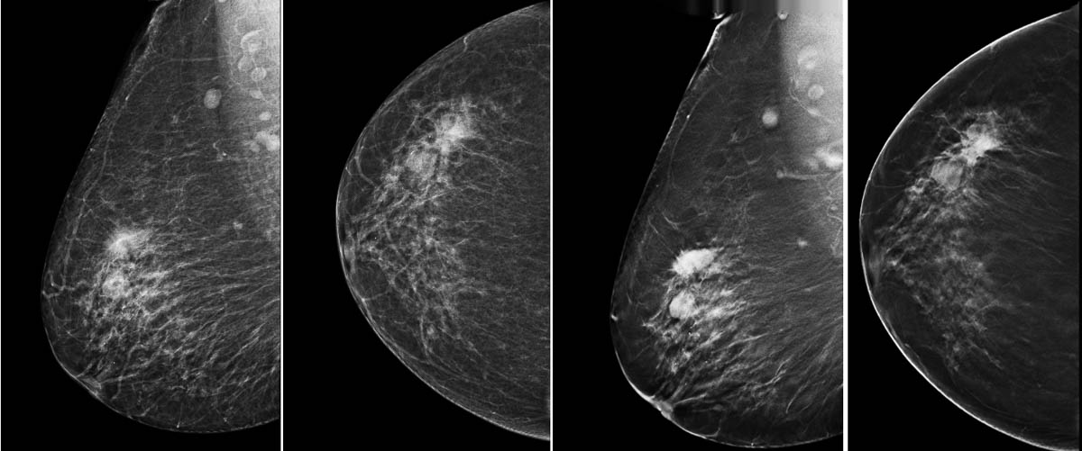
L'intérêt de la tomosynthèse est d'éliminer les superpositions glandulaires mammaires, ce qui améliore les performances diagnostiques de sensibilité et de spécificité pour la détection des cancers du sein.
Dépistage du cancer du sein
Après une revue critique de la littérature, le volet 1 du rapport de la Haute Autorité de santé (HAS) sur la performance de la mammographie par tomosynthèse dans le dépistage organisé du cancer du sei

Discussion
Aucun commentaire
Commenter cet article