Introduction

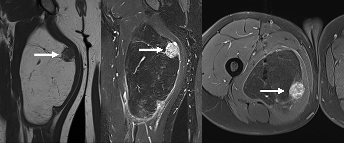
Les tumeurs des tissus mous représentent une gamme hétérogène de lésions bénignes ou malignes. Ces lésions sont regroupées selon différentes catégories, incluant les tumeurs adipeuses (lipomes, liposarcomes), les tumeurs fibroblastiques (tumeurs desmoides…), les tumeurs des gaines nerveuses périphériques (schwannome, tumeur maligne des gaines nerveuses périphériques…), etc. De nombreuses formations peuvent également simuler un aspect de tumeur (« pseudo-tumeurs »), par...

Mickael TORDJMAN

Médecin radiologue

Hôpital Raymond-Poincaré – Assistance publique-Hôpitaux de Paris

Garches

Bibliographie
  1. Honoré C., Méeus P., Stoeckle E. et coll., « Soft tissue sarcoma in France in 2015: Epidemiology, classification and organization of clinical care », Journal of Visceral Surgery, septembre 2015, vol. 152, n° 4, p. 223-230. DOI : 10.1016/j.jviscsurg.2015.05.001.
  2. WHO classification of tumours; soft tissue and bone tumours, 5e édition, volume 3, IARC Press, 2020.
  3. National Cancer Institute, « National Cancer Institute, DCCPS, Surveillance Research Program, Cancer Statistics Branch; Surveillance, Epidemiology, and End Results (SEER) Program », SEER Database: Incidence - SEER Cancer Statistics Review 1975-2017. https://seer.cancer.gov/data/. Site consulté le 20 août 2021.
  4. Crombé A., Marcellin P.-J., Buy X. et coll., « Soft tissue sarcomas: assessment of MRI features correlating with histologic grade and patient outcome », Radiology, avril 2019, vol. 291, n° 3, p. 710-721. DOI : 10.1148/radiol.2019181659.
  5. Noebauer-Huhmann I. M., Weber M.-A., Lalam R. K. et coll., « Soft tissue tumors in adults: ESSR-approved guidelines for diagnostic imaging », Seminars in musculoskeletal radiology, décembre 2015, vol. 19, n° 5, p. 475-482. DOI : 10.1055/s-0035-1569251.
  6. Kransdorf M. J. et Murphey M. D., Imaging of Soft Tissue Tumors, 3e édition, Philadelphia, Wolters Kluwet Health - Lippincott Williams & Wilkins, 2013, 776 p.
  7. Wilkerson B. W., Crim J. R., Hung M. et coll., « Characterization of synovial sarcoma calcification », American Journal of Roentgenology, décembre 2012, vol. 199, n° 6, p. 730-734. DOI : 10.2214/AJR.11.7342.
  8. Wortman J. R., Tirumani S. H., Jagannathan J. P. et coll., « Primary extremity liposarcoma : MRI features, histopathology, and clinical outcomes », Journal of Computer Assisted Tomography, septembre-octobre 2016, vol. 40, n° 5, p. 791-798. DOI : 10.1097/RCT.0000000000000431.
  9. Kransdorf J. M., Bancroft L. W., Peterson J. J. et coll., « Imaging of fatty tumors: distinction of lipoma and well-differentiated liposarcoma », Radiology, juillet 2002, vol. 224, n° 1, p. 99-104. DOI : 10.1148/radiol.2241011113.
  10. Rizer M., Singer A. D., Edgar M. et coll., « The histological variants of liposarcoma : predictive MRI findings with prognostic implications, management, follow-up, and differential diagnosis », Skeletal Radiology, septembre 2016, vol. 45, n° 9, p. 1193-1204. DOI : 10.1007/s00256-016-2409-4. Epub : 21 mai 2016.
  11. Kawai A., Woodruf J., Healey J. H. et coll., « SYT-SSX gene fusion as a determinant of morphology and prognosis in synovial sarcoma », New England Journal of Medicine, janvier 1998, vol. 338, n° 3, p. 153‑160. DOI : 10.1056/NEJM199801153380303.
  12. Trassard M., Le Doussal V., Hacène K. et coll., « Prognostic factors in localized primary synovial sarcoma: a multicenter study of 128 adult patients », Journal of Clinical Oncology, janvier 2001, vol. 19, n° 2, p. 525‑534. DOI : 10.1200/JCO.2001.19.2.525.
  13. Murphey M. D., Gibson M. S., Jennings B. T. et coll., « Imaging of synovial sarcoma with radiologic-pathologic correlation », RadioGraphics, septembre 2006, vol. 26, n° 5, p. 1543‑1565. DOI : 10.1148/rg.265065084.
  14. Domagoj A. V., Spelman T., May D. et coll., « Predicting the prognosis of undifferentiated pleomorphic soft tissue sarcoma: a 20-year experience of 266 cases », ANZ Journal of Surgery, septembre 2019, vol. 89, n° 9, p. 1045-1050. DOI : 10.1111/ans.15348.
  15. Murphey M. D., Gross T. M., Rosenthal H. G., « Musculoskeletal malignant fibrous histiocytoma: radiologic-pathologic correlation », RadioGraphics, juillet 1994, vol. 14, n° 4, p. 807‑826. DOI : 10.1148/radiographics.14.4.7938770.
  16. Weiss A. R., Lyden E. R., Anderson J. R. et coll., « Histologic and clinical characteristics can guide staging evaluations for children and adolescents with rhabdomyosarcoma: a report from the Children’s Oncology Group Soft Tissue Sarcoma Committee », Journal of Clinical Oncology, septembre 2013, vol. 31, n° 26, p. 3226-3232. DOI : 10.1200/JCO.2012.44.6476. Epub : 12 août 2013.
  17. Inarejos Clemente E. J., Navallas M., Barber Martínez de la Torre I. et coll., « MRI of rhabdomyosarcoma and other soft-tissue sarcomas in children », Radiographics, mai 2020, vol. 40, n° 3, p. 791-814. DOI : 10.1148/rg.2020190119.
  18. James A. W., Shurell E., Singh A. et coll., « Malignant peripheral nerve sheath tumor », Surgical Oncology Clinics of North America, octobre 2016, vol. 25, n° 4, p. 789-802. DOI : 10.1016/j.soc.2016.05.009.
  19. Wasa J., Nishida Y., Tsukushi S. et coll., « MRI features in the differentiation of malignant peripheral nerve sheath tumors and neurofibromas », American Journal of Roentgenology, juin 2010, vol. 194, n° 6, p. 1568-1574. DOI : 10.2214/AJR.09.2724.