Imagerie du foie

Évaluation de la stéatose hépatique en imagerie

icon réservé aux abonnésArticle réservé aux abonnés
Marco Dioguardi Burgio Le 07/07/20 à 7:00, mise à jour hier à 15:05 Lecture 13 min.

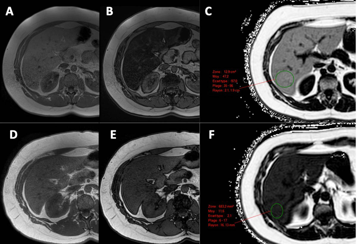
L’IRM, avec la mesure de la Proton Density Fat Fraction (PDFF), est considérée aujourd’hui comme l’examen de référence pour quantifier la stéatose hépatique. © Marco Dioguardi Burgio

Résumé

L’utilisation de méthodes d’imagerie pour la quantification non invasive de la stéatose hépatique devient de plus en plus récurrente dans la pratique clinique. Les différentes méthodes d’imagerie (échographie, scanner et IRM), peuvent être utilisées en fonction de leur disponibilité et du contexte clinique. De plus, pour chaque modalité d’imagerie, différents types d’approche (évaluation semi-quantitative ou quantitative) peuvent être appliqués pour réaliser cette quantification. Si l’IRM avec la mesure de la Proton Density Fat Fraction (PDFF) est considérée aujourd’hui comme l’examen de référence, l’échographie reste la modalité la plus utilisée dans la pratique clinique.

Introduction

La réalisation d’examens d’imagerie pour la quantification non invasive de la stéatose hépatique devient de plus en plus récurrente dans la pratique clinique. L’incidence de la maladie du foie gras non alcoolique (non alcoholic fatty liver disease – NAFLD) a augmenté au cours des dernières décennies et sa prévalence mondiale actuelle est d’environ 30 % [1]. La NAFLD est associée à d’autres conditions pathologiques comme l’obésité, l’hypertension artérielle, le diabète de type 2 et le syndrome métabolique [2, 3]. De plus, la NAFLD peut se compliquer d’une atteinte inflammatoire. Cette forme inflammatoire, la stéatohépatite non alcoolique (non alcoholic steatohepatitis – NASH), peut évoluer vers la cirrhose et ses complications (notamment l’hypertension portale et le carcinome hépatocellulaire) [1, 4, 5]. Parallèlement à la quantification de la stéatose hépatique, l'imagerie joue un rôle important dans l'évaluation non invasive de l'inflammation et de la fibrose hépatique che

Il vous reste 94% de l’article à lire

Docteur Imago réserve cet article à ses abonnés

S'abonner à l'édition
  • Tous les contenus « abonnés » en illimité
  • Le journal numérique en avant-première
  • Newsletters exclusives, club abonnés

Abonnez-vous !

Docteur Imago en illimité sur desktop, tablette, smartphone, une offre 100% numérique

Offre mensuelle 100 % numérique

23 €

par mois

S’abonner à Docteur Imago

Auteurs

Marco Dioguardi Burgio

Radiologue Hôpital Beaujon Clichy

Bibliographie

  1. Younossi Z. M., Koenig A. B., Abdelatif D. et coll., « Global epidemiology of nonalcoholic fatty liver disease-Meta-analytic assessment of prevalence, incidence, and outcomes », Hepatology, juillet 2016, vol. 64, n° 1, p. 73-84. DOI : 10.1002/hep.28431.
  2. Ballestri S., Zona S., Targher G., et coll., « Nonalcoholic fatty liver disease is associated with an almost twofold increased risk of incident type 2 diabetes and metabolic syndrome. Evidence from a systematic review and meta-analysis », Journal of Gastroenterology and Hepatology, mai 2016, vol. 31, n° 5, p. 936-944. DOI : 10.1111/jgh.13264.
  3. Targher G., Byrne C. D., Lonardo A., « Non-alcoholic fatty liver disease and risk of incident cardiovascular disease: A meta-analysis », Journal of Hepatology, septembre 2016, vol. 65, n° 3, p. 589-600. DOI : 10.1016/j.jhep.2016.05.013.
  4. Ekstedt M., Franzén L. E., Mathiesen U. L. et coll., « Long-term follow-up of patients with NAFLD and elevated liver enzymes », Hepatology, octobre 2006, vol. 44, n° 4, p. 865-873. DOI : 10.1002/hep.21327.
  5. EASL, EASD, EASO, « Clinical practice guidelines for the management of non-alcoholic fatty liver disease », Journal of Hepatology, juin 2016, vol. 64, n° 6, p. 1388-1402. DOI : 10.1016/j.jhep.2015.11.004.
  6. Adinolfi L. E., Gambardella M., Andreana A. et coll., « Steatosis accelerates the progression of liver damage of chronic hepatitis C patients and correlates with specific HCV genotype and visceral obesity », Hepatology, juin 2001, vol. 33, n° 6, p. 1358-1364. DOI : 10.1053/jhep.2001.24432.
  7. McCormack L., Petrowsky H., Jochum W. et coll., « Hepatic steatosis is a risk factor for postoperative complications after major hepatectomy: a matched case-control study », Annals of Surgery, juin 2007, vol. 245, n° 6, p. 923-930. DOI : 10.1097%2F01.sla.0000251747.80025.b7.
  8. Pais R., Barritt A. S., Calmus Y. et coll., « NAFLD and liver transplantation: Current burden and expected challenges », Journal of Hepatology, décembre 2016, vol. 65, n° 6, p. 1245-1257. DOI : 10.1016/j.jhep.2016.07.033.
  9. Brunt E. M., Janney C. G., Di Bisceglie A. M. et coll., « Nonalcoholic steatohepatitis: a proposal for grading and staging the histological lesions », The American Journal of Gastroenterology, septembre 1999, vol. 94, n° 9, p. 2467-2474. DOI : 10.1016/S0002-9270(99)00433-5.
  10. Strauss S., Gavish E., Gottlieb P. et coll., « Interobserver and intraobserver variability in the sonographic assessment of fatty liver », AJR American Journal of Roentgenology, décembre 2007, vol. 189, n° 6, p. W320-323. DOI : 10.2214/AJR.07.2123.
  11. Bohte A. E., van Werven J. R., Bipat S. et coll., « The diagnostic accuracy of US, CT, MRI and 1H-MRS for the evaluation of hepatic steatosis compared with liver biopsy: a meta-analysis », European Radiology, janvier 2011, vol. 21, n° 1, p. 87-97. DOI : 10.1007%2Fs00330-010-1905-5.
  12. Chevallier P., « Qu’attendre de l’imagerie médicale non invasive pour dépister et quantifier la stéatose hépatique ? », Gastroentérologie Clinique et Biologique, novembre 2005, vol. 29, n° 11, p. 1133-1135. DOI : GCB-11-2005-29-11-0399-8320-101019-200516267.
  13. Marshall R. H., Eissa M., Bluth E. I. et coll., « Hepatorenal index as an accurate, simple, and effective tool in screening for steatosis », AJR American Journal of Roentgenology, novembre 2012, vol. 199, n° 5, p. 997-1002. DOI : 10.2214/AJR.11.6677.
  14. Chauhan A., Sultan L. R., Furth E. E. et coll., « Diagnostic accuracy of hepatorenal index in the detection and grading of hepatic steatosis », Journal of Clinical Ultrasound : JCU, novembre 2016, vol. 44, n° 9, p. 580-586. DOI : 10.1002/jcu.22382.
  15. Bae J. S., Lee D. H., Lee J. Y. et coll., « Assessment of hepatic steatosis by using attenuation imaging: a quantitative, easy-to-perform ultrasound technique », European Radiology, décembre 2019, vol. 29, n° 12, p. 6499-6507. DOI : 10.1007/s00330-019-06272-y.
  16. Dioguardi Burgio M., Ronot M., Reizine E. et coll., « Quantification of hepatic steatosis with ultrasound: promising role of attenuation imaging coefficient in a biopsy-proven cohort », European Radiology, avril 2020, vol. 30, n° 4, p. 2293-2301. DOI : 10.1007/s00330-019-06480-6.
  17. Dioguardi Burgio M., Imbault M., Ronot M. et coll., « Ultrasonic adaptive sound speed estimation for the diagnosis and quantification of hepatic steatosis: a pilot study », Ultraschall in der Medizin, novembre 2018, vol. 40, n° 60. DOI : 10.1055/a-0660-9465.
  18. Han A., Zhang Y. N., Boehringer A. S. et coll., « Assessment of hepatic steatosis in nonalcoholic fatty liver disease by using quantitative US », Radiology, avril 2020, vol. 295, n° 1, p. 106-113. DOI : 10.1148/radiol.2020191152.
  19. Piekarski J., Goldberg H. I., Royal S. A., « Difference between liver and spleen CT numbers in the normal adult: its usefulness in predicting the presence of diffuse liver disease », Radiology, décembre 1980, vol. 137, n° 3, p. 727-729. DOI : 10.1148/radiology.137.3.6934563.
  20. Fischer M. A., Gnannt R., Raptis D A. et coll., « Quantification of liver fat in the presence of iron and iodine : an ex-vivo dual-energy CT study », Investigative Radiology, juin 2011, vol. 46, n° 6, p. 351-358. DOI : 10.1097/RLI.0b013e31820e1486.
  21. Hyodo T., Yada N., Hori M., et coll., « Multimaterial decomposition algorithm for the quantification of liver fat content by using fast-kilovolt-peak switching dual-energy CT: clinical evaluation », Radiology, avril 2017, vol. 283, n° 1, p. 108-118. DOI : 10.1148/radiol.2017160130.
  22. Kodama Y., Ng C. S., Wu T. T. et coll., « Comparison of CT methods for determining the fat content of the liver », AJR American Journal of Roentgenology, mai 2007, vol. 188, n° 5, p. 1307-1312. DOI : 10.2214/AJR.06.0992.
  23. Szczepaniak L. S., Nurenberg P., Leonard D., et coll., « Magnetic resonance spectroscopy to measure hepatic triglyceride content: prevalence of hepatic steatosis in the general population », American Journal of Physiology. Endocrinology and Metabolism, février 2005, vol. 288, n° 2, E462-468. DOI : 10.1152/ajpendo.00064.2004.
  24. Lv S., Jiang S., Liu S. et coll., « Noninvasive quantitative detection methods of liver fat content in nonalcoholic fatty liver disease », Journal of Clinical and Translational Hepatology, juin 2018, vol. 6, n° 2. p. 217-221. DOI : 10.14218/JCTH.2018.00021.
  25. Stern C., Castera L., « Non-invasive diagnosis of hepatic steatosis », Hepatology International, janvier 2017, vol. 11, n° 1, p. 70-78. DOI : 0.1007/s12072-016-9772-z.
  26. Qu Y., Li M., Hamilton G. et coll., « Diagnostic accuracy of hepatic proton density fat fraction measured by magnetic resonance imaging for the evaluation of liver steatosis with histology as reference standard: a meta-analysis », European Radiology, octobre 2019, vol. 29, n° 10, p. 5180-5189. DOI : 10.1007/s00330-019-06071-5.

Discussion

Aucun commentaire

Laisser un commentaire

Sur le même thème

Le fil Docteur Imago

11 Août

7:20

Dans une étude mettant en avant les disparités dans le dépistage complémentaire des femmes ayant des seins denses, les chercheurs soulignent la nécessité de standardiser l'évaluation de la densité mammaire, de renforcer la formation des professionnels et d'investir dans les infrastructures afin d'améliorer la détection précoce du cancer du sein.
10 Août

14:00

Les coûts croissants et les inefficacités opérationnelles menacent la durabilité financière de l’IRM. L’optimisation des flux de travail, de la formation, de la planification et l’intégration de l’IA peuvent améliorer l’efficacité et générer des gains de revenus estimés entre 40 et 60 %, selon des projections rapportées dans une étude de Clinical Imaging.

12:10

07 Août

16:00

Pour la détection du cancer du sein, les performances diagnostiques des protocoles d'IRM abrégée par rapport à l'IRM standard varient selon le protocole et le contexte clinique. La technique FAST conserve une sensibilité élevée, tandis que la combinaison FAST + ultrafast + T2W offre une spécificité supérieure dans un contexte diagnostique (étude).
Docteur Imago

GRATUIT
VOIR