Introduction

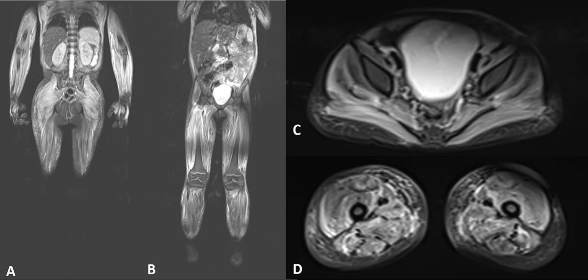
L’IRM corps entier est un examen particulièrement attractif en imagerie pédiatrique. Elle fournit une vision d’ensemble du corps humain en un seul examen, permettant de ne pas multiplier les explorations. Elle n’expose pas aux rayonnements ionisants et répond donc aux principes fondamentaux de la radioprotection. Le principe ALARA, « As Low As Reasonably Achievable », est ainsi respecté. L’IRM corps entier constitue donc une alternative intéressante aux autres modalités...

Inès MANNES

Radiologue

Service d'imagerie pédiatrique

Hôpital Bicêtre - Assistance publique - Hôpitaux de Paris

Bibliographie
  1. Laor T., Jaramillo D., « MR Imaging Insights into Skeletal Maturation: What Is Normal ? », Radiology, janvier 2009, vol. 250, n° 1, p. 28–38. DOI : 10.1148/radiol.2501071322. Article en libre accès.
  2. Raissaki M., Demetriou S., Spanakis K. et coll., « Multifocal Bone and Bone Marrow Lesions in Children — MRI findings », Pediatric Radiology, mars 2017, vol. 47, n° 3, p. 342–360. DOI : 10.1007/s00247-016-3737-1.
  3. Greer M.-L. C., « Whole-body Magnetic Resonance Imaging: Techniques and Non-Oncologic Indications », Pediatric Radiology, août 2018, vol. 48, n° 9, p. 1348–1363. DOI : 10.1007/s00247-018-4141-9.
  4. Falip C., Alison M., Boutry N. et coll., « Chronic Recurrent Multifocal Osteomyelitis (CRMO): A Longitudinal Case Series Review », Pediatric Radiology, mars 2013, vol. 43, n° 3, p. 355–375. DOI : 10.1007/s00247-012-2544-6.
  5. Fritz J., Tzaribatchev N., Claussen C. D. et coll., « Chronic Recurrent Multifocal Osteomyelitis: Comparison of Whole-Body MR Imaging with Radiography and Correlation with Clinical and Laboratory Data », Radiology, septembre 2009, vol. 252, n° 3, p. 842–851. DOI : 10.1148/radiol.2523081335. Article en libre accès.
  6. Voit A. M., Arnoldi A. P., Douis H. et coll., « Whole-body Magnetic Resonance Imaging in Chronic Recurrent Multifocal Osteomyelitis: Clinical Longterm Assessment May Underestimate Activity », The Journal of Rheumatology, août 2015, vol. 42, n° 8, p. 1455–1462. DOI : 10.3899/jrheum.141026.
  7. Eutsler E. P., Khanna G., « Whole-body magnetic resonance imaging in children: technique and clinical applications », Pediatric Radiology, 26 mai 2016, vol. 46, p. 858–872. DOI : 10.1007/s00247-016-3586-y.
  8. Huang Z.-G., Gao B. X., Chen H. et coll., « An efficacy analysis of whole-body magnetic resonance imaging in the diagnosis and follow-up of polymyositis and dermatomyositis », PloS One, 17 juin 2017, vol. 12, n° 17, e0181069. DOI : 10.1371/journal.pone.0181069.
  9. Lecouvet F. E., « Whole-Body MR Imaging: Musculoskeletal Applications », Radiology, mai 2016, vol. 279, n° 2, p. 345–365. DOI : 10.1148/radiol.2016142084. Article en libre accès.
  10. Kim J. R., Yoon H. M., Jung A. Y. et coll., « Comparison of whole-body MRI, bone scan, and radiographic skeletal survey for lesion detection and risk stratification of Langerhans Cell Histiocytosis », Scientific Reports, 2019, vol. 9, n° 317. DOI : 10.1038/s41598-018-36501-1. Article en libre accès.
  11. Goo H. W., Yang D. H., Ra Y. S., et coll., « Whole-body MRI of Langerhans cell histiocytosis: comparison with radiography and bone scintigraphy », Pediatric Radiology, octobre 2006, vol. 36, p. 1019–1031. DOI : 10.1007/s00247-006-0246-7.
  12. Perez-Rossello J. M., Connolly S. A., Newton A. W., et coll., « Whole-body MRI in Suspected Infant Abuse », AJR American Journal of Roentgenology, septembre 2010, vol. 195, n° 3, p. 744–750. DOI : 10.2214/AJR.09.3364.
  13. Davis, J. T., Kwatra, N., Schooler, G. R., « Pediatric whole-body MRI: A review of current imaging techniques and clinical applications », Journal of Magnetic Resonance Imaging, 4 avril 2016, vol. 44, n° 4, p. 783–793. DOI : 10.1002/jmri.25259.
  14. Gottumukkala, R. V., Gee, M. S., Hampilos, P. J. et coll., « Current and Emerging Roles of Whole-Body MRI in Evaluation of Pediatric Cancer Patients », RadioGraphics, 2019, vol. 39, n° 2, p. 516-534. DOI : 10.1148/rg.2019180130.