Le terme de conflit de Haglund, maladie de Haglund, etc., est souvent évoqué en cas de douleur ou de tuméfaction de la zone d’insertion basse du tendon d’Achille et tout le monde fait référence à la description princeps du suédois Patrick Haglund en 1928 [1]. Les descriptions de Haglund concernaient des déformations de l’extrémité postérosupérieure du calcanéus et des tuméfactions des tissus mous adjacents ; il se basait sur l’observation de certains de ses patients, nobles autrichiens, dont les escarpins plats, à tige postérieure rigide, favorisaient un conflit à la face postérieure du calcanéus [2].
En fait, ce qui est important est de ne pas confondre la cause de la lésion et ses conséquences.
La cause est bien un conflit entre le « coin » postérosupérieur du calcaneus et la face antérieure du tendon d’Achille distal. Ce conflit entraîne une dilacération parfois fissuraire des fibres antérieures du tendon et parfois une bursite réactionnelle. Celle-ci est un épiphénomène et c’est sur
Pathologies des membres inférieurs
Le conflit de Haglund : causes et conséquences
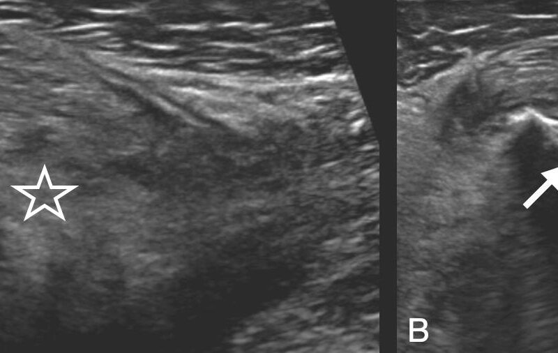
Le conflit de Haglund est un conflit entre le « coin » postérosupérieur du calcaneus et la face antérieure du tendon d’Achille distal, qui entraîne une dilacération parfois fissuraire des fibres antérieures du tendon et parfois une bursite réactionnelle. Il survient notamment chez les patients présentant une proéminence anormale du « coin » postérosupérieur du calcaneus, une verticalisation du calcaneus, ou une déformation de l’arrière-pied entraînant une pression asymétrique. Une hyperutilisation ou encore le port de chaussures plates avec des renforts postérieurs rigides sont également des facteurs étiologiques. L’imagerie est utilisée pour détecter le conflit, évaluer la gravité de la dilacération tendineuse et rechercher les facteurs favorisants. Les clichés de radiologie standards sont indispensables pour évaluer la verticalisation calcanéenne, mesurer l’arche plantaire à la recherche d’un pied creux, la morphologie du coin postérocrânial du calcanéus, l’infiltration du triangle graisseux de Kager et la présence de calcifications et d’ossifications. L’échographie intervient en seconde intention pour réaliser un examen comparatif en tension puis en position détendue. L’IRM présente un aspect cartographique de l’ensemble des lésions très utile en particulier en préopératoire. Elle montre l’importance de la lésion tendineuse ainsi que, chez certains patients, un hypersignal T2 du calcanéus postérieur confirmant le conflit ; les fissures sont souvent bien précisées en IRM, à condition qu’elles ne soient pas collabées par une tension trop importante lors de l’installation du patient. Le traitement doit en premier lieu agir sur les causes de cette lésion mécanique primitive plutôt que sur les atteintes inflammatoires secondaires.
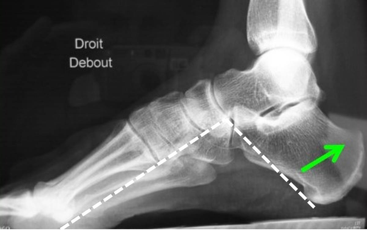
Les clichés de radiographie sont incontournables et permettent notamment de déterminer l’obliquité du calcanéus par rapport au sol, sa verticalisation étant l'une des causes du conflit de Haglund. © Brasseur J.-L.
Il vous reste 97% de l’article à lire
Docteur Imago réserve cet article à ses abonnés
Vous avez déjà un compte ? Se connecter

- Tous les contenus « abonnés » en illimité
- Le journal numérique en avant-première
- Newsletters exclusives, club abonnés
Abonnez-vous !
Docteur Imago en illimité sur desktop, tablette, smartphone, une offre 100% numérique
23 €
par mois
Auteurs
Jean-Louis Brasseur
Radiologue Service de radiologie polyvalente et oncologique, AP-HP Paris
Insep
Paris
Imagerie médicale de la Plaine de France
Montfermeil
Bibliographie
- Haglund P., « Breitrag zur Kliniken des Achilessehne », Zeitschrift für Orthopadie und Chirurgie, 1928, vol. 49, p. 4-7.
- Peetrons P., Vanderhofstadt A., « La maladie de Haglund revisitée » in Brasseur J.-L., Zeitoun-Eiss D., Renoux J. et coll., Actualités en échographie de l’appareil locomoteur (Tome 4), Sauramps Médical, Montpellier, 2007, p. 163-173.
- Kang S., Thordarson D. B., Charlton T. P., « Insertional Achilles tendinitis and Haglund’s deformity », Foot & Ankle International, juin 2012, vol. 33, n° 6, p. 487-491. DOI : 10.3113/FAI.2012.0487.
- Reinherz R. P., Smith B. A., Henning K. E., « Understanding the pathologic Haglund’s deformity », The Journal of Foot Surgery, septembre-octobre 1990, vol. 29, n° 5, p. 432-435. PMID : 2258561.
- Stephens M. M., « Haglund’s deformity and retrocalcaneal bursitis », The Orthopedic Clinics of North America, janvier 1994, vol. 25, n° 1, p. 41-46. PMID : 8290230.
- Vaishya R., Agarwal A. K., Azizi A. T. et coll., « Haglund’s Syndrome: A Commonly Seen Mysterious Condition », Cureus, octobre 2016, vol. 8, n° 10 : e820. DOI : 10.7759/cureus.820.
- Pavlov H., Heneghan M. A., Hersh A. et coll., « The Haglund syndrome: initial and differential diagnosis », Radiology, juillet 1982, vol. 144, n° 1, p. 83-88. DOI : 10.1148/radiology.144.1.7089270.
- Kucuksen S., Karahan A. Y., Erol K., « Haglund syndrome with pump bump », Medical Archives, 2012, vol. 66, n° 6, p. 425-427. DOI : 10.5455/medarh.2012.66.425-427.
- Van Dijk C. N., van Sterkenburg M. N., Wiegerinck J. I. et coll., « Terminology for Achilles tendon related disorders », Knee Surgery, Sports Traumatology, Arthroscopy, 2011, vol. 19, n° 5, p. 835-841. DOI : 10.1007/s00167-010-1374-z. Epub 11 janvier 2011.
- Lohrer H., Arentz S., [Impingement lesion of the distal anterior Achilles tendon in sub-Achilles bursitis and Haglund-pseudoexostosis-a therapeutic challenge], Sportverletz Sportschaden, décembre 2003, vol. 17, n° 4, p. 181-188. DOI : 10.1055/s-2003-45406.
- Lohrer H., Nauck T., « Retrocalcaneal bursitis but not Achilles tendinopathy is characterized by increased pressure in the retrocalcaneal bursa », Clinical Biomechanics (Bristol, Avon), mars 2014, vol. 29, n° 3, p. 283-288. DOI : 10.1016/j.clinbiomech.2013.12.002. Epub 9 décembre 2013.
- Morvan G., Vuillemin V., Guerini H. et coll., « Le tendon d’Achille. De la biomécanique à l’imagerie », in Brasseur J.-L., Mercy G., Massein A. et coll., Actualités en échographie de l’appareil locomoteur (Tome 11), Sauramps Médical, Montpellier, 2014, p. 11-32.
- Benjamin M., Toumi H., Ralphs J.-R. et coll., « Where tendons and ligaments meet bone: attachment sites (‘entheses’) in relation to exercise and/or mechanical load », Journal of Anatomy, avril 2006, vol. 208, n° 4, p. 471-490. DOI : 10.1111/j.1469-7580.2006.00540.x.
- Milz S., Rufai A., Buettner A. et coll., « Three-dimensional reconstructions of the Achilles tendon insertion in man », Journal of Anatomy, février 2002, vol. 200, n° 2, p. 145-152. DOI : 10.1046%2Fj.0021-8782.2001.00016.x.
- Theobald P., Bydder G., Dent C. et coll., « The functional anatomy of Kager’s fat pad in relation to retrocalcaneal problems and other hindfoot disorders », Journal of Anatomy, janvier 2006, vol. 208, n° 1, p. 91-97. DOI : 10.1111/j.1469-7580.2006.00510.x.
- Shaw H. M., Vásquez O. T., McGonagle D. et coll., « Development of the human Achilles tendon enthesis organ », Journal of Anatomy, décembre 2008, vol. 213, n° 6, p. 718-724. DOI : 10.1111%2Fj.1469-7580.2008.00997.x.
- Oegema T. R., Carpenter R. J., Hofmeister F. et coll., « The interaction of the zone of calcified cartilage and subchondral bone in osteoarthritis », Microscopy Research and Technique, mai 1997, vol. 37, n° 4, p. 324-332. DOI : 10.1002/(sici)1097-0029(19970515)37:4%3C324::aid-jemt7%3E3.0.co;2-k.
- Kim P. J., Richey J.-M., Wissman L. R. et coll., « The variability of the Achilles tendon insertion: a cadaveric examination », The Journal of Foot and Ankle Surgery, septembre – octobre 2010, vol. 49, n° 5, p. 417-420. DOI : 10.1053/j.jfas.2010.05.002. Epub 25 juin 2010.
- Ballal M. S., Walker C. R., Molloy A. P., « The anatomical footprint of the Achilles tendon: a cadaveric study », The Bone & Joint Journal, octobre 2014, vol. 96-B, n° 10, p. 1344-1348. DOI : 10.1302/0301-620X.96B10.33771.
- Mahan J., Damodar D., Trapana E. et coll., « Achilles tendon complex: The anatomy of its insertional footprint on the calcaneus and clinical implications », Journal of Orthopaedics, juin 2019, vol. 17, p. 221-227. DOI : 10.1016/j.jor.2019.06.008.
- Edama M., Kubo M., Onishi H. et coll., « Structure of the Achilles tendon at the insertion on the calcaneal tuberosity », Journal of Anatomy, novembre 2016, vol. 229, n° 5, p. 610-614. DOI : 10.1111/joa.12514. Epub 22 juin 2016.
- Shaw H. M., Santer R. M., Watson A. H. D. et coll., « Adipose tissue at entheses: the innervation and cell composition of the retromalleolar fat pad associated with the rat Achilles tendon », Journal of Anatomy, octobre 2007, vol. 211, n° 4, p. 436-443. DOI : 10.1111%2Fj.1469-7580.2007.00791.x.
- Court-Payen M., Cardinal E., Dakhil Delfi A. et coll., « Lésions distales du tendon calcanéen » in Morvan G., Bianchi S., Bouysset M. et coll. (dir), Le pied, Sauramps Médical, Montpellier, 2011, 331-344.
- Porsch M., Hackenbroch M. H., König D. P., [Atypical Achilles tendon rupture in Haglund exostosis–a case report], Zeitschrift fur Orthopadie und Ihre Grenzgebiete, novembre-décembre 1998, vol. 136, n° 6, p. 568-570. DOI : 10.1055/s-2008-1045188.
- Morel M., Boutry N., Demondion X. et coll., « Normal anatomy of the heel entheses: anatomical and ultrasonographic study of their blood supply », Surgical and Radiologic Anatomy : SRA, août 2005, vol. 27, n° 3, p. 176-183. DOI : 10.1007/s00276-004-0311-6. Epub 26 mai 2005.
- Chauveaux D., Liet P., Lehuec J. C. et coll., « A new radiologic measurement for the diagnosis of Haglund’s deformity », Surgical and Radiologic Anatomy : SRA, 1991, vol. 13, n° 1, p. 39-44. DOI : 10.1007/bf01623140.
- Bulstra G. H., van Rheenen T. A., Scholtes V. A. B., « Can we measure the heel bump? Radiographic evaluation of Haglund’s deformity », The Journal of Foot and Ankle Surgery, mai-juin 2015, vol. 54, n° 3, p. 338-340. DOI : 10.1053/j.jfas.2014.07.006. Epub 30 août 2014.
- Tourné Y., Baray A.-L., Barthélémy R. et coll., « Contribution of a new radiologic calcaneal measurement to the treatment decision tree in Haglund syndrome », Orthopaedics & Traumatology, Surgery & Research, décembre 2018, vol. 104, n° 8, p. 1215-1219. DOI : 10.1016/j.otsr.2018.08.014. Epub 31 octobre 2018.
- Morvan G., Brasseur J.-L., Échographie de la cheville et du pied, Sauramps Médical, Montpellier, 2012, 332 p.
- Bianchi S., Martinoli C., Ultrasound of the musculoskeletal system, Springer Berlin Heidelberg, New-York, 2007, 978 p.
- Bullock M. J., Mourelatos J., Mar A., « Achilles impingement tendinopathy on magnetic resonance imaging », The Journal of Foot and Ankle Surgery, mai-juin 2017, vol. 56, n° 3, p. 555-563. DOI : 10.1053/j.jfas.2017.01.024.
- Kouvalchouk J.-F., Watin-Augouard L., « Chirurgie du tendon d’Achille : tendinites et maladies de Haglund » in EMC – Techniques chirurgicales orthopédie-traumatologie, Elsevier, Paris, 1993 : 1-4, 44-912.
- Xu J. H., Ding S.-L., Chen B. et coll., « Modified Bunnell suture expands the surgical indication of the treatment of Haglund’s syndrome heel pain with endoscope », Experimental and Therapeutic Medicine, juin 2018, vol. 15, n° 6, p. 4817-4821. DOI : 10.3892/etm.2018.6071. Epub 13 avril 2018.
- Maynou C., Mestdagh H., Dubois H. H. et coll., « L’ostéotomie calcanéenne est-elle justifiée dans la maladie de Haglund ? », Revue de chirurgie orthopédique, 1998, vol. 84, n° 8, p. 734-738.
- Hardy A., Rousseau R., Issa S.-P. et coll., « Functional outcomes and return to sports after surgical treatment of insertional Achilles tendinopathy: Surgical approach tailored to the degree of tendon involvement », Orthopaedics & Traumatology, Surgery & Research, septembre 2018, vol. 104, n° 5, p. 719-723. DOI : 10.1016/j.otsr.2018.05.003. Epub 28 mai 2018.
- Brunner J., Anderson J., O’Malley M. et coll., « Physician and patient based outcomes following surgical resection of Haglund’s deformity », Acta Orthopaedica Belgica, décembre 2005, vol. 71, n° 6, p. 718-723. PMID : 16459864.
Derniers articles de la rubrique IRM

Derniers articles de la rubrique Échographie

Derniers articles de la rubrique Radiologie conventionnelle
Derniers articles de la rubrique Musculo-squelettique
Discussion
Aucun commentaire
Commenter cet article