Contexte
La nécessité de définir une prise en charge adaptée
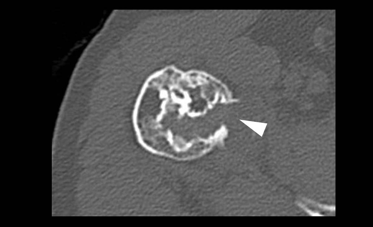
Les lésions osseuses focales (LOF) sont fréquentes, mais il y a une grande disparité de prévalence entre les tumeurs bénignes et pseudo-lésions (très fréquentes) et les tumeurs malignes (rares) [1, 2]. Le problème est que les tumeurs osseuses malignes ou bénignes mais localement agressives nécessitent une prise en charge particulière, passant souvent par une chirurgie carcinologique qui doit être réalisée dans un centre expert...

- Rajiah P., Ilaslan H., Sundaram M., « Imaging of primary malignant bone tumors (nonhematological) », Radiology Clinics of North America, novembre 2011, vol. 49, n° 6, p. 1135‑1161, v. Epub : 21 septembre 2011. DOI : 10.1016/j.rcl.2011.07.003.
- Plant J., Cannon S., « Diagnostic work up and recognition of primary bone tumours: a review », EFORT Open Reviews, juin 2016, vol. 1, n° 6, p. 247‑253. Epub : 8 juin 2016. DOI : 10.1302/2058-5241.1.000035.
- Casali P. G., Bielack S., Abecassis N. et coll., « Bone sarcomas: ESMO-PaedCan-EURACAN clinical practice guidelines for diagnosis, treatment and follow-up », Annals of Oncology : Official Journal of the European Society for Medical Oncology, octobre 2018, vol. 29 (Suppl 4), p. iv79‑ iv95. DOI : 10.1093/annonc/mdy310.
- Choi J. H., Ro J. Y., « The 2020 WHO classification of tumors of bone: an updated review », Advances in Anatomic Pathology, mai 2021, vol. 28, n° 3, p. 119‑138. DOI : 10.1097/PAP.0000000000000293.
- Miller T. T., « Bone tumors and tumorlike conditions: analysis with conventional radiography », Radiology, mars 2008, vol. 246, n° 3, p. 662‑674. Epub : 25 janvier 2008. DOI : 10.1148/radiol.2463061038.
- Guillin R., Moser T., Koob M. et coll., « Subperiosteal hematoma of the iliac bone: imaging features of acute and chronic stages with emphasis on pathophysiology », Skeletal Radiology, juin 2012, vol. 41, n° 6, p. 667‑675. Epub : 14 septembre 2011. DOI : 10.1007/s00256-011-1267-3.
- Madewell J. E., Ragsdale B. D., Sweet D. E., « Radiologic and pathologic analysis of solitary bone lesions. Part I: internal margins », Radiologic Clinics of North America, décembre 1981, vol. 19, n° 4, p. 715‑748. PMID : 7323290.
- Ragsdale B. D., Madewell J. E., Sweet D. E., « Radiologic and pathologic analysis of solitary bone lesions. Part II: periosteal reactions », Radiologic Clinics of North America, décembre 1981, vol. 19, n° 4, p. 749‑783. PMID : 7323291.
- Sweet D. E., Madewell J. E., Ragsdale B. D., « Radiologic and pathologic analysis of solitary bone lesions. Part III: matrix patterns », Radiologic Clinics of North America, décembre 1981, vol. 19, n° 4, p. 785‑814. PMID : 7323292.
- Do B. H., Langlotz C., Beaulieu C. F., « Bone tumor diagnosis using a naïve bayesian model of demographic and radiographic features », Journal of Digital Imaging, octobre 2017, vol. 30, n° 5, p. 640‑647. DOI : 10.1007/s10278-017-0001-7.
- Expert panel on musculoskeletal imaging, Bestic J. M., Wessell D. E., Beaman F. D. et coll., « ACR Appropriateness Criteria® Primary Bone Tumors », Journal of the American College of Radiology : JACR, mai 2020, vol. 17, n° 5S, p. S226‑S238. DOI : https://doi.org/10.1016/j.jacr.2020.01.038.
- Tang H., Ahlawat S., Fayad L. M., « Multiparametric MR imaging of benign and malignant bone lesions », Magnetic Resonance Imaging Clinics in North America, novembre 2018, vol. 26, n° 4, p. 559‑569. DOI : 10.1016/j.mric.2018.06.010.
- Ribeiro G. J., Gillet R., Hossu G. et coll., « Solitary bone tumor imaging reporting and data system (BTI-RADS): initial assessment of a systematic imaging evaluation and comprehensive reporting method », European Radiology, octobre 2021, vol. 31, n° 10, p. 7637-7652. Epub : 25 mars 2021. DOI : 10.1007/s00330-021-07745-9.