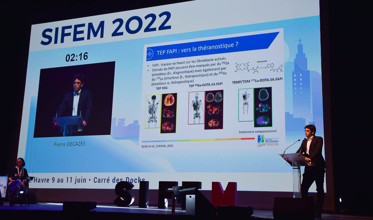
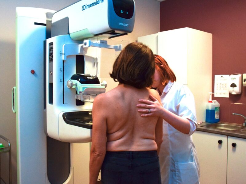
Le 10 juin dernier, le congrès 2022 de la Société d’imagerie de la femme (SIFEM), a dédié une journée à l’imagerie mammaire. Pierre Decazes, médecin nucléaire au centre Henri-Becquerel, à Rouen (76), a évoqué le potentiel de l’imagerie TEP pour le bilan du cancer du sein. À l’heure actuelle, la TEP au fluorodésoxyglucose (FDG) est présente dans toutes les recommandations internationales, rappelle-t-il. En France, les recommandations de l’Institut national du cancer, publiées en octobre 2021, la réservent au bilan à distance. « On ne fait pas de TEP pour le bilan local ou ganglionnaire. En revanche, c’est intéressant pour la stadification des cancers de stades cliniques IIA et IIB, et au-delà », précise Pierre Decazes.
Sensibilité et spécificité limitées dans le bilan local
Pour ce qui est du bilan local, la TEP au FDG n’est donc pas indiquée pour la caractérisation lésionnelle. « Sa sensibilité est limitée, remarque l’intervenant. Pour les lésions de taille inférieure ou égale à 10 mm, i


Discussion
Aucun commentaire
Commenter cet article