Automatisation en radiologie interventionnelle

Robotique endovasculaire et intelligence artificielle : un avenir prometteur !

icon réservé aux abonnésArticle réservé aux abonnés
Tom Boeken Le 06/05/25 à 15:00 Lecture 13 min.

Résumé

La combinaison de l’intelligence artificielle et de la robotique transformera à moyen terme l’exercice de la radiologie interventionnelle. L’IA rendra l’image intelligente et intelligible pour le robot en fournissant le support technologique pour réaliser l’acte manuel de manière plus autonome. Les clés de cette évolution résident dans le traitement des images endovasculaires en temps réel pour lequel plusieurs solutions ont fait leurs preuves, notamment en neuroradiologie et en imagerie abdominale. Le suivi en direct du matériel à l’écran associé aux cathéters intelligents permettra en parallèle d’avancer vers l’autonomie au bloc de RI.

Introduction : intelligence artificielle et radiologie interventionnelle

Computer vision et robotique vers l’autonomie au bloc

L’intelligence artificielle (IA) est déjà bien ancrée dans la pratique de la radiologie diagnostique. L’IA, issue du monde du gaming et des statistiques, a également de grandes chances d’impacter la radiologie interventionnelle.

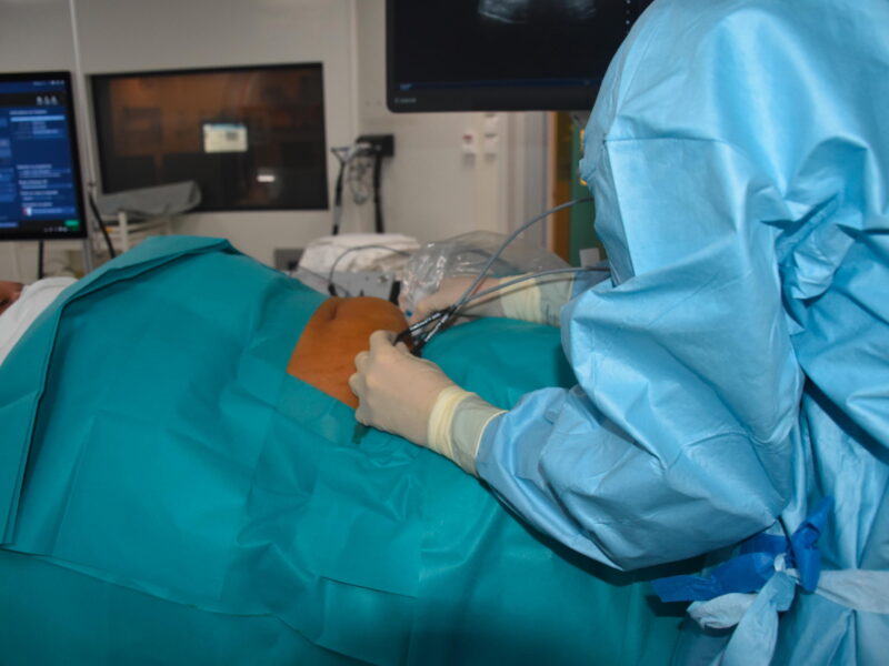
Le radiologue interventionnel utilise l’image pour se guider et ses mains pour manipuler des dispositifs dans le patient. Ces deux facultés, l’une intellectuelle et l’autre manuelle, sont au cœur de multiples innovations. D’un côté la computer vision ou « compréhension » de l’image, de l’autre la robotique. En alliant les deux, nous pouvons imaginer approcher l’autonomie au bloc où l’humain n’aura pas tout à fait le même rôle.

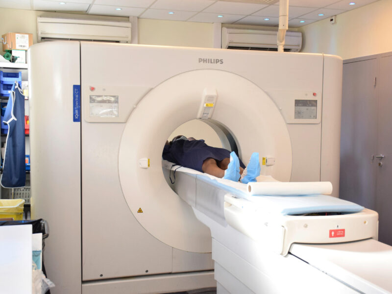
L’IA s’installe en interventionnel pour le diagnostic

La computer vision est la branche des statistiques qui s’intéresse à la « compréhension » de l’image. En réalité, les termes « compréhension » ou « intelligence » f

Il vous reste 95% de l’article à lire

Docteur Imago réserve cet article à ses abonnés

S'abonner à l'édition
  • Tous les contenus « abonnés » en illimité
  • Le journal numérique en avant-première
  • Newsletters exclusives, club abonnés

Abonnez-vous !

Docteur Imago en illimité sur desktop, tablette, smartphone, une offre 100% numérique

Offre mensuelle 100 % numérique

23 €

par mois

S’abonner à Docteur Imago

Auteurs

Tom Boeken

MCU-PH Service de radiologie interventionnelle vasculaire et oncologique, Hôpital européen Georges-Pompidou, laboratoire HeKA, INRIA Paris

Bibliographie

  1. Boeken T, Feydy J, Lecler A, et al (2023) Artificial intelligence in diagnostic and interventional radiology : Where are we now ? Diagnostic and Interventional Imaging 104 : 1 – 5. https://doi.org/10.1016/j.diii.2022.11.004.
  2. Boeken T, Feydy J, Lecler A, et al (2023) Artificial intelligence in diagnostic and interventional radiology : Where are we now ? Diagnostic and Interventional Imaging 104 : 1 – 5. https://doi.org/10.1016/j.diii.2022.11.004.
  3. Hughes H, Cornelis FH, Scaglione M, Patlas MN (2025) Paranoid About Androids : A Review of Robotics in Radiology. Can Assoc Radiol J 76 : 232 – 238. https://doi.org/10.1177/08465371241290076.
  4. Milot L, Soyer P (2025) Robotics in Interventional Radiology : Is the Force With Us ? Can Assoc Radiol J 76 : 199 – 200. https://doi.org/10.1177/08465371241299645.
  5. Solomon SB, Patriciu A, Bohlman ME, et al (2002) Robotically Driven Interventions : A Method of Using CT Fluoroscopy without Radiation Exposure to the Physician. Radiology 225 : 277 – 282. https://doi.org/10.1148/radiol.2251011133.
  6. Rafii-Tari H, Payne CJ, Yang G-Z (2014) Current and Emerging Robot-Assisted Endovascular Catheterization Technologies : A Review. Ann Biomed Eng 42 : 697 – 715. https://doi.org/10.1007/s10439-013-0946-8.
  7. Weisz G, Metzger DC, Caputo RP, et al (2013) Safety and Feasibility of Robotic Percutaneous Coronary Intervention. Journal of the American College of Cardiology 61 : 1596 – 1600. https://doi.org/10.1016/j.jacc.2012.12.045.
  8. Mahmud E, Schmid F, Kalmar P, et al (2016) Feasibility and Safety of Robotic Peripheral Vascular Interventions. JACC : Cardiovascular Interventions 9 : 2058 – 2064. https://doi.org/10.1016/j.jcin.2016.07.002.
  9. Mendes Pereira V, Cancelliere NM, Nicholson P, et al (2020) First-in-human, robotic-assisted neuroendovascular intervention. J NeuroIntervent Surg 12 : 338 – 340. https://doi.org/10.1136/neurintsurg-2019-015671.rep.
  10. Von Zur Mühlen C, Jeuck M, Heidt T, et al (2024) One-year outcome of robotical vs. manual percutaneous coronary intervention. Clin Res Cardiol. https://doi.org/10.1007/s00392-024-02524-0.
  11. Shekhtman O, Sioutas GS, Dagli MM, et al (2024) Robotic spinal angiography : A single-center experience. Interv Neuroradiol 15910199241272515. https://doi.org/10.1177/15910199241272515.
  12. Mendes Pereira V, Rice H, De Villiers L, et al (2024) Evaluation of effectiveness and safety of the CorPath GRX robotic system in endovascular embolization procedures of cerebral aneurysms. J NeuroIntervent Surg 16 : 405 – 411. https://doi.org/10.1136/jnis-2023-020161.
  13. Durand E, Sabatier R, Smits PC, et al (2023) Evaluation of the R-One robotic system for percutaneous coronary intervention : the R-EVOLUTION study. EuroIntervention 18 : e1339 – e1347. https://doi.org/10.4244/EIJ-D-22-00642.
  14. Vidal V, Bargellini I, Bent C, et al (2024) Performance Evaluation of a Miniature and Disposable Endovascular Robotic Device. Cardiovasc Intervent Radiol 47 : 503 – 507. https://doi.org/10.1007/s00270-024-03686-0.
  15. (2024) First Use of the Sentante Endovascular Robotic System in Human Patients – Sentante. https://sentante.com/first-use-of-the-sentante-endovascular-robotic-system-in-human-patients/. Consulté le 28 mars 2025.
  16. Ghosh R, Wong K, Zhang YJ, et al (2024) Automated catheter segmentation and tip detection in cerebral angiography with topology-aware geometric deep learning. J NeuroIntervent Surg 16 : 290 – 295. https://doi.org/10.1136/jnis-2023-020300.
  17. Sakakura Y, Kono K, Fujimoto T (2025) Real time artificial intelligence assisted carotid artery stenting : a preliminary experience. J NeuroIntervent Surg 17 : 410 – 414. https://doi.org/10.1136/jnis-2024-021600.
  18. Klaassen J, Hazenberg CEVB, Bloemert-Tuin T, et al (2024) Editor’s Choice – Radiation Dose Reduction During Contralateral Limb Cannulation Using Fiber Optic RealShape Technology in Endovascular Aneurysm Repair. European Journal of Vascular and Endovascular Surgery 67 : 594 – 600. https://doi.org/10.1016/j.ejvs.2023.10.041.

Discussion

Aucun commentaire

Laisser un commentaire

Sur le même thème

Le fil Docteur Imago

17 Avr

14:33

Une revue systématique publiée dans Radiography conclut au fort potentiel de l’utilisation du jumeau numérique en IRM, notamment en cardiologie et en oncologie, malgré des limites dans les domaines de la formation, de la sécurité et de l’intégration opérationnelle.

7:12

Une étude publiée dans Radiography analyse l’utilisation du modèle d’apprentissage profond Att-U-Net pour segmenter les tissus pulmonaires et les tumeurs à partir d’images PET-CT, afin d’améliorer le diagnostic du cancer du poumon. Les résultats montrent de bonnes performances (DSC 0,81 et IoU 0,69), suggérant que ce modèle pourrait renforcer la précision clinique et faciliter la planification des traitements.
16 Avr

15:41

Mount Sinai est le premier au monde à utiliser le système TheraSphere™ Y-90 « Any Day Dosing », un traitement mini-invasif qui délivre directement des radiations aux tumeurs du foie via le sang. Cette innovation permet de traiter les patients plus rapidement et plus souvent dans la semaine, améliorant ainsi l’accès aux soins et réduisant les délais, annonce un communiqué. 

13:16

Une revue systématique et méta-analyse démontre que la mammographie avec contraste (CEM) présente une très forte valeur prédictive négative pour les asymétries non rehaussées, avec un risque de cancer extrêmement faible. En revanche, la présence de rehaussement est fortement associée à la malignité et permet d’améliorer la stratification du risque.

7:30

Une étude aux Pays-Bas montre qu’un protocole standardisé améliore partiellement la qualité des comptes rendus d’échographie thyroïdienne, mais reste inégalement appliqué. Son efficacité dépend surtout d’une bonne communication et collaboration entre les professionnels de santé.

Docteur Imago

GRATUIT
VOIR