Le 29/07/26 à 14:17
Le score LI-RADS v2024 sans rayonnements a démontré d'excellentes performances diagnostiques et s'est révélé performant pour évaluer la viabilité tumorale chez des patients atteints d'un CHC traités par radiothérapie faible dose associée à une immunothérapie ciblée (
étude).
Le 29/07/26 à 7:47
Les adolescents sportifs présenteraient des différences spécifiques à la région, à la zone et à la couche dans les temps de relaxation du cartilage du genou T2 par rapport aux non sportifs. Cette cartographie pourrait permettre de détecter les cartilages à risques avant que des dommages structurels visibles ne se produisent. (
Etude)
Le 28/07/26 à 15:00
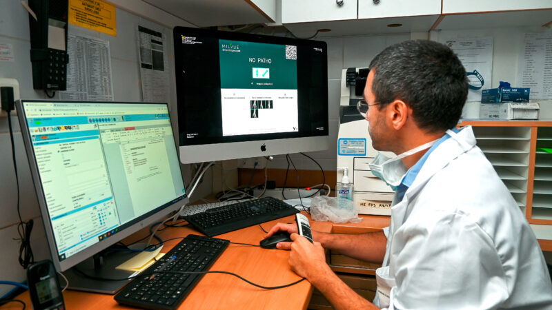
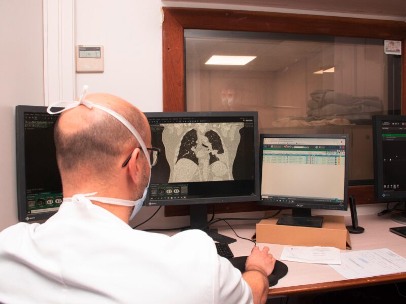
Dans
une étude présentée dans Clinical Radiology, des radiologues ne sont parvenus à identifier comme telles que 75 % d'images générées par intelligence artificielle. Des résultats proches de ceux
d'une autre étude sur le même sujet.
Le 28/07/26 à 11:00
Un score incluant des variables extraites des examens baseline de TEP-TDM au [
18F]FDG et au [
68Ga]Ga-PSMA-11 offre des informations utiles pour le pronostic des patients atteints de cancer de la prostate résistant à la castration pour lesquels un traitement au [
177Lu]Lu-PSMA-617 est envisagé, conclut
une étude française.
Le 28/07/26 à 7:30
Dans une étude observationnelle menée chez des femmes aux seins denses, l'utilisation de l'échographie automatique du sein (ABUS) a été associée à un taux moins important de résultats anormaux et de recommandations de suivi à court-terme que l'utilisation de l'échographie manuelle, pour un taux de détection des cancers similaire.

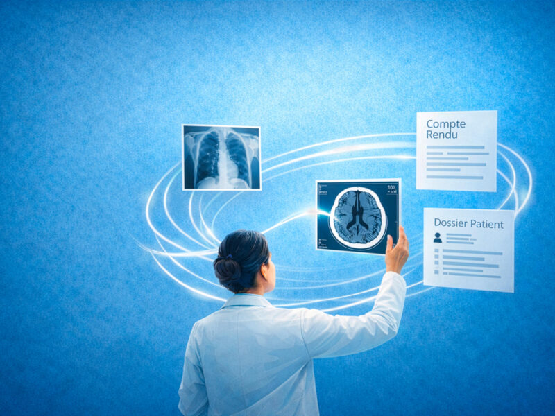
 Les recommandations de l’Académie de médecine pour maîtriser les dangers et limites de l’IA en imagerie